 education
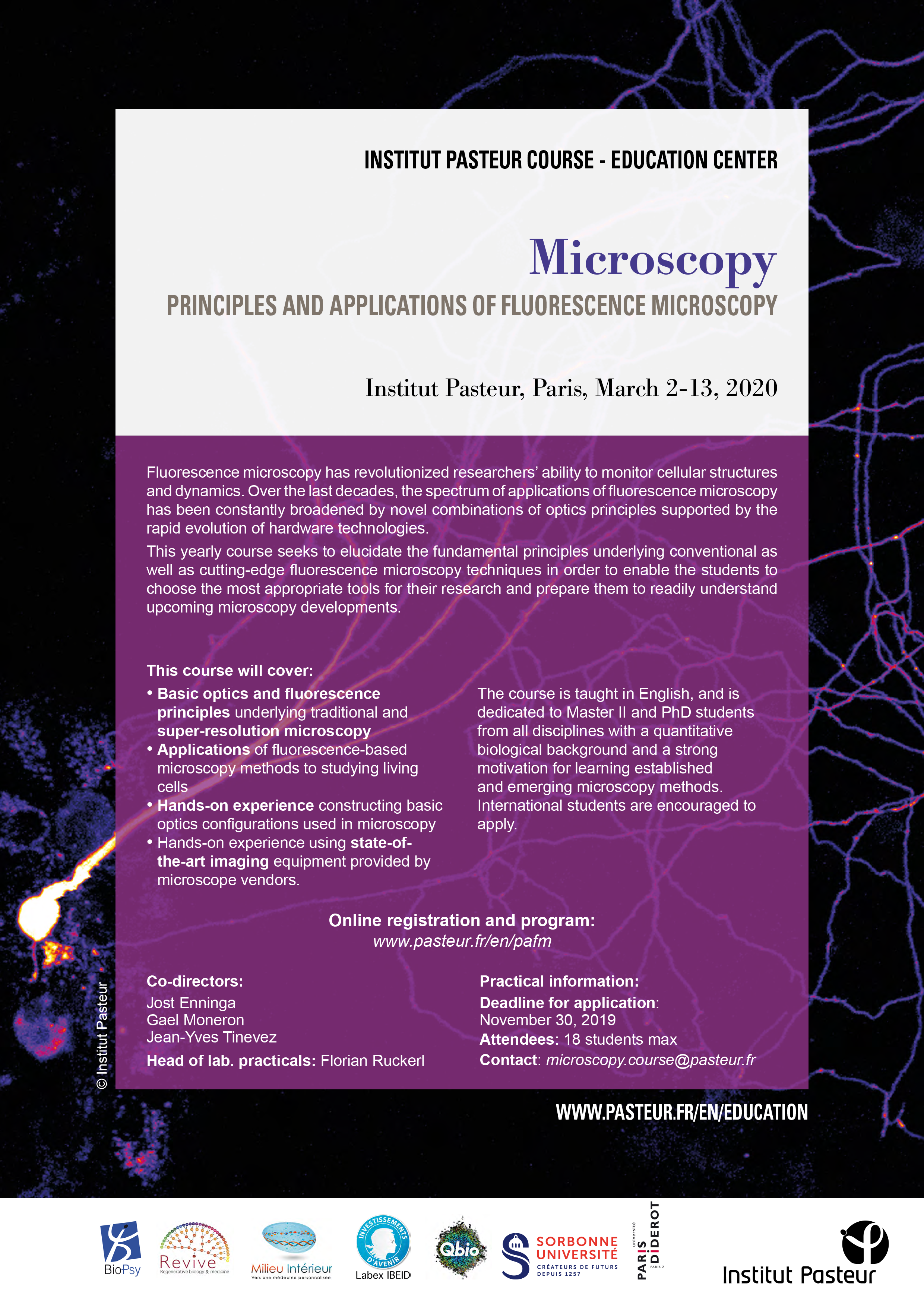
Extension of submission deadline for the "Principles and Applications of Fluorescence Microscopy" course
 The use of fluorescence microscopy (wide field, confocal, multiphoton, and now superresolution) in combination with genetically encoded fluorescence probes comprise a powerful set of scientific tools to study live cells. However, surprising little practical and theoretical training in such methods exists within standard curricula, particular at the early stage of training (Masters or Doctorate level).
This course offers to cover basic optics principles necessary to understand the origin of microscope resolution and design. Participants will get hands-on experience implementing simple optical configurations to illustrate these fundamental principles. Subsequently, participants will perform experiments on state-of-the-art imaging equipment provided by microscope vendors.
This course (March 2 to March 13, 2020) is intended for Master’s students and international PhD students who expect to be using advanced live cell fluorescence microscopy for future studies. The registration date for this course has been extended. You now have until November 30 to register.
Lectures will cover, in depth, the principle behind traditional high resolution imaging methods such as confocal, multi-photon, and the recently developed super-resolutions methods. Students will also learn about fundamental properties of synthetic and genetically encoded fluorescence indicators used for cellular morphology imaging and signaling recording. Finally guest lectures will demonstrate applications of some of the discussed methodologies.