 Museum
Museum collections: artifacts from the early days of photography! 12 photomicrographs dating from 1867 currently being restored
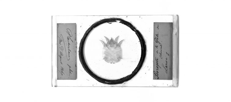
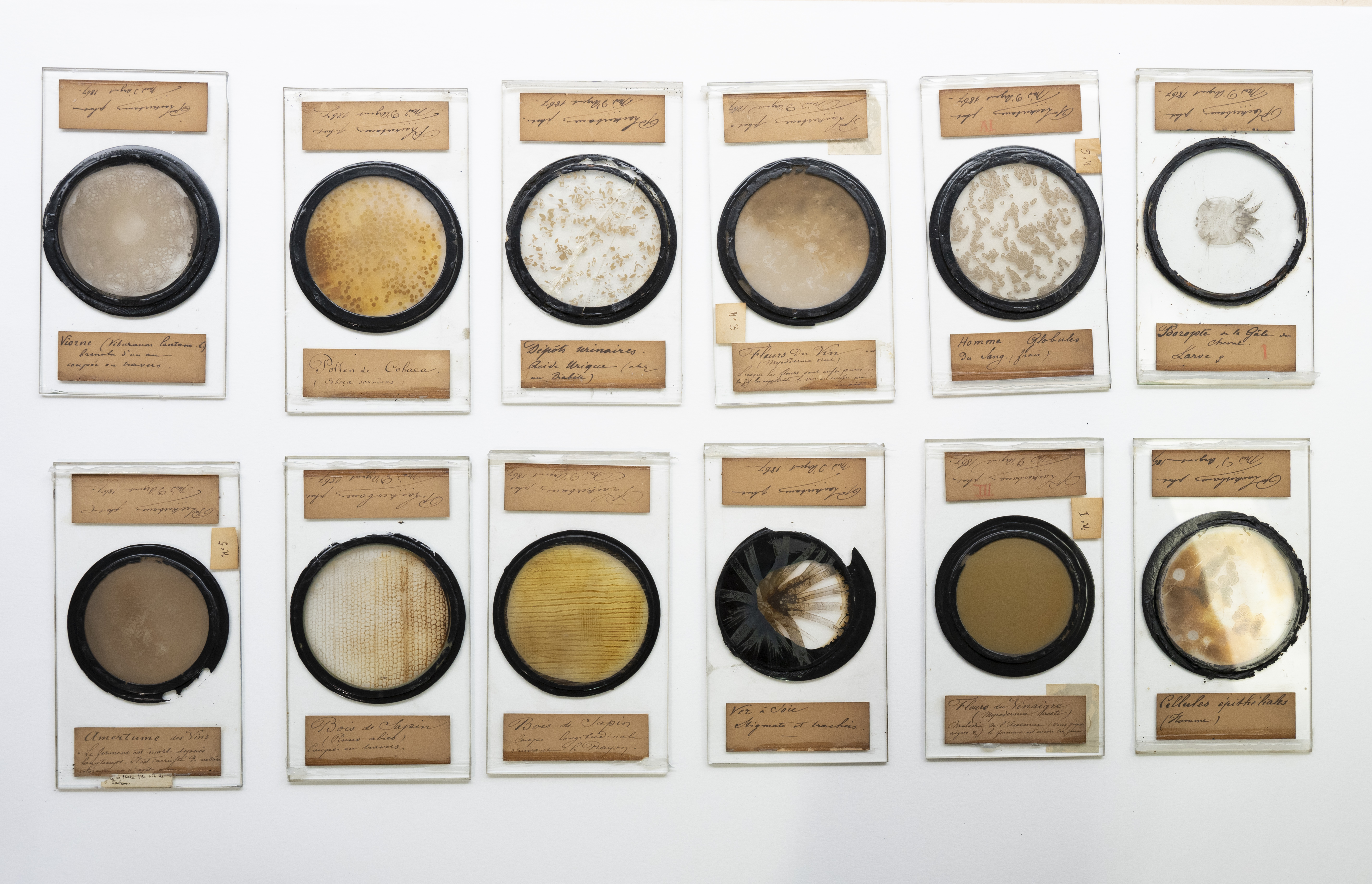
A few months ago, the Pasteur Museum rediscovered twelve photomicrographs on glass plates dating from 1867. Previously on display in the former Museum of Research Applications in Marnes-la-Coquette, the photomicrographs bear witness to the early history of photography as used to study the invisible living world.
These photographic plates were produced with wet collodion, a technique developed by Frederick Scott Archer (1813-1857) from 1851 onwards. The process was widely used until the 1880s, when it was replaced with more stable gelatine-silver bromide emulsions.
![]()
 What is the wet-collodion photographic process?
Collodion is a nitrocellulose solution dissolved in a mix of ether and alcohol. It was discovered by Louis Ménard (1822-1901) in 1846. The wet-collodion photographic process involves applying a collodion emulsion to a glass plate. To make the collodion light sensitive, soluble iodides and bromides are added. Once the plate is coated, it is immersed in a silver nitrate bath, making it photosensitive.
The technique has a major drawback: the negative has to be prepared, exposed and then developed in a very short space of time, as once it dries it stops being sensitive and is impossible to develop. Depending on the temperature and humidity conditions, the entire process must be completed in no longer than 15 to 30 minutes.
![]()
These photomicrographs were produced when Louis Pasteur was finishing his research on wine, for which he received the Grand Prix at the Universal Exposition on July 1, 1867. At the time he was also researching silkworm diseases. This was also the period when he completed three years of scientific teaching at the École des Beaux-Arts in Paris.
These glass plates bear witness to this point in Louis Pasteur's scientific career and his insatiable curiosity. On the labels of the glass plates, which are still clearly legible, we read: pine wood (Pinus abies), viburnum (Viburnum lantan), Cobaea pollen (Cobaea scandens), human epithelial cells, red blood cells, urine sample from an individual with diabetes, silkworm stigmata and tracheae, bitterness of wines, vinegar flowers (Mycoderma aceti) and Psoroptes larva, a mite species responsible for equine scabies.
The photomicrographs of vinegar flowers were reproduced in Louis Pasteur's book Etudes sur le Vinaigre (1868).
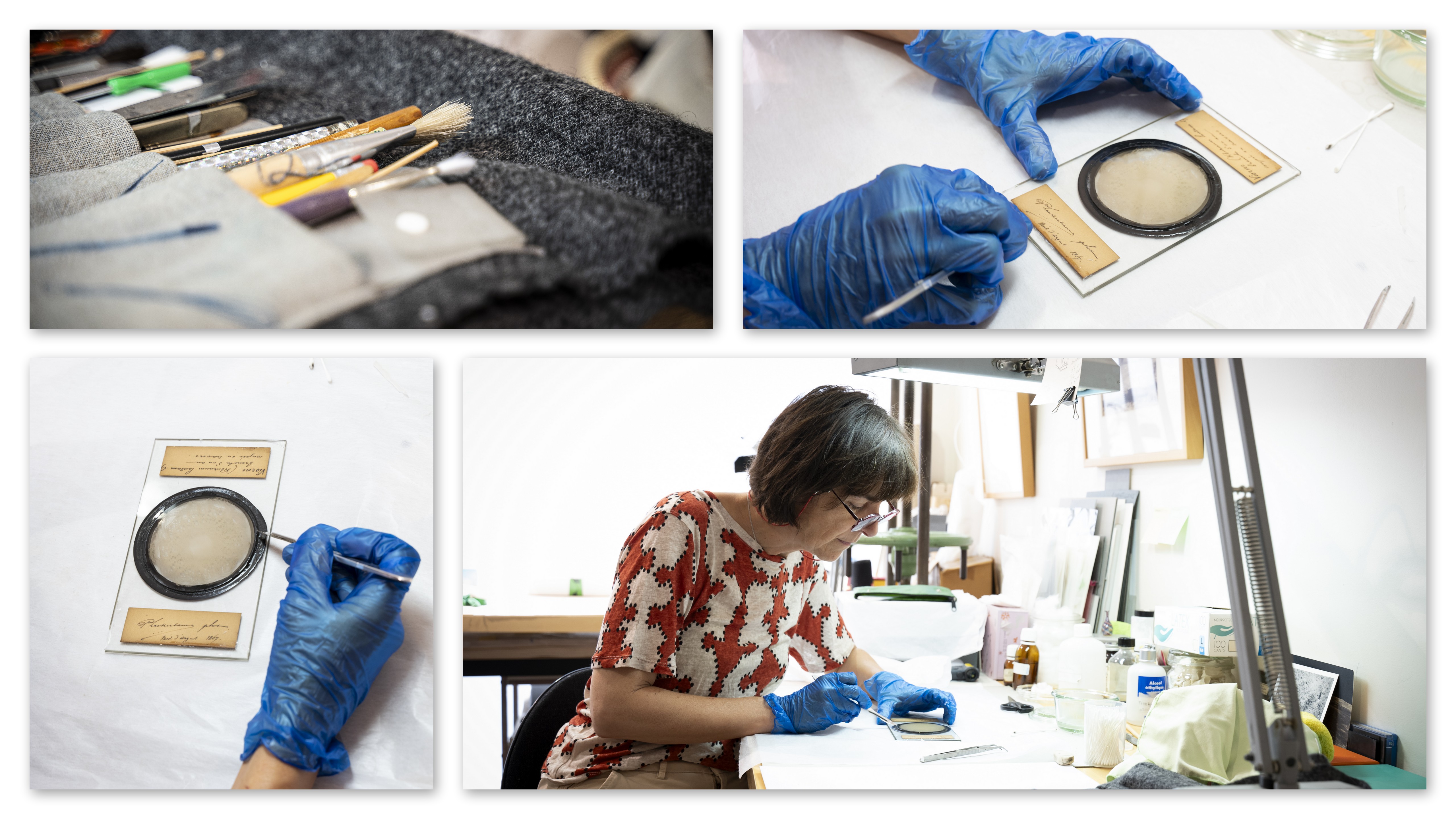
Estelle Rebourt, a restorer of historical photos, is currently working on these precious glass plates to clean them and strengthen the setting that fixes the image to the glass plate. The museum team has contacted the Research and Restoration Center for French Museums (C2RMF) to determine the nature of the black compound used to fix the image.
Watch the restoration video (in French)
![]()
 What is the difference between a photomicrograph and a microphotograph?
Photomicrography refers to all the photographic techniques used to photograph a subject with the help of a microscope. Emile Roux was a pioneer in this technique, founding the first photomicrography laboratory at the Institut Pasteur in 1889.
Microphotography, invented by René Dagron (1819-1900), involves producing very small images (1mm in diameter). Microphotography was widely used in espionage techniques.
Photos: François Gardy/Institut Pasteur

