May 02, 2025
Bulletin interne de l'Institut Pasteur
Condition report exercise with students from the National Heritage Institute: photomicrography
On April 3 and 4, the Pasteur Museum hosted a group of curation and restoration students from the National Heritage Institute (INP), together with Antonin Riou, a specialist photograph restorer, to carry out a condition report in the museum's storage facility.

The INP students and the museum team in the storage facility
Two students in photography restoration and two students in archive curation from the National Heritage Institute (INP), as well as a curator from the National Museum of the Democratic Republic of the Congo, came to carry out an assignment in the museum storage facility. Their task was to produce a condition report for the photomicrography collection. The aim was to examine nearly 200 items and determine any damage (mechanical, chemical or biological). The students then issued recommendations for reconditioning these photomicrographs and potentially including them in the museum's permanent collection in 2028.
This practical exercise was an opportunity for discussions between students and museum professionals. The students were able to put their knowledge into practice in a real-world setting with a fieldwork exercise. Hosting the students for this assignment also gave the museum team a better understanding of this collection and taught us more about the photography techniques used in laboratories in the 19th and early 20th centuries.
Photographing invisible living worlds: photomicrography
Photomicrography, not to be confused with microphotography, refers to a series of techniques used to photograph subjects using a microscope. More often than not, scientists produce these shots by replacing the eyepiece of the microscope with a lens and removing the camera lens so that the microscope can form an image on the camera sensor.
The photomicrographs in the Pasteur Museum collections are key historical documents that shine a light on the history of life sciences.
Émile Roux (1853-1933) was a pioneer in photomicrography. In 1887 he published an article in the Annales de l'Institut Pasteur in which he gave a precise description of a photomicrographic apparatus that is now part of the museum's collections. Émile Roux also founded the Institut Pasteur's first photomicrography laboratory in 1889. In 1903, Paul Jeantet took over as head of the laboratory, continuing the experimental work using this technique.
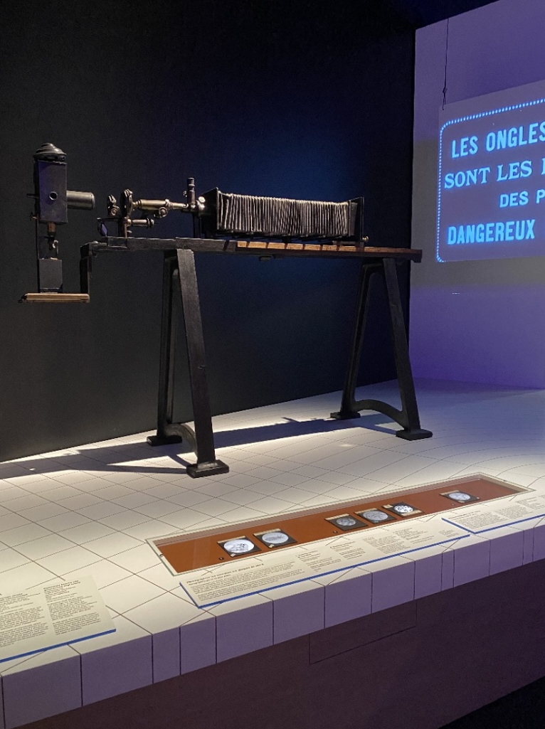
Photomicrographic apparatus belonging to Émile Roux presented at the exhibition "Epidemics. Taking care of the living world" at the Musée des Confluences in 2024.
The museum has photomicrography specimens produced using several different techniques:
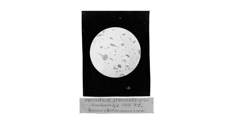
1) Photomicrographs produced using wet collodion on glass plates
Collodion is a nitrocellulose solution dissolved in a mix of ether and alcohol. It was discovered by Louis Ménard (1822-1901) in 1846. The wet-collodion photographic process involves applying a collodion emulsion to a glass plate. To make the collodion light sensitive, soluble iodides and bromides are added. Once the plate is coated, it is immersed in a silver nitrate bath, making it photosensitive.
The technique has a major drawback: the negative has to be prepared, exposed and then developed in a very short space of time, as once it dries it stops being sensitive and is impossible to develop. Depending on the temperature and humidity conditions, the entire process must be completed in no longer than 15 to 30 minutes.
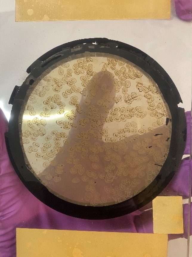
Photomicrographs produced using wet collodion on glass plates
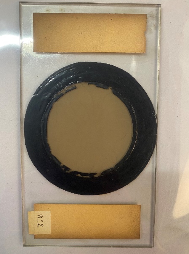
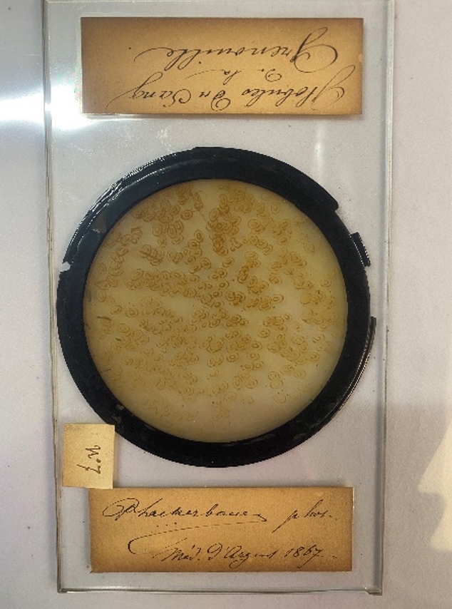
2) Photomicrographs produced using gelatin silver printing
The gelatin silver process, invented in 1871 by Richard Leach Maddox (1816-1902), is a chemical process used in analog photography. A suspension of silver halides (or silver salts) in gelatin is applied to a glass plate, plastic film or baryta paper.
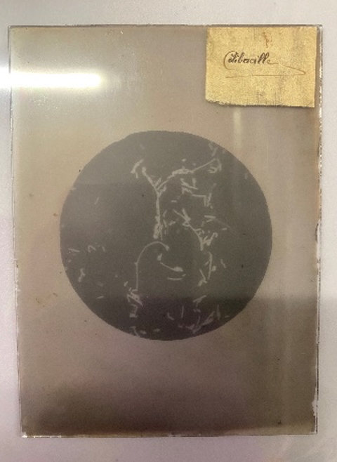
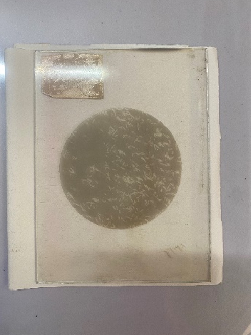
Photomicrographs – gelatin silver positives on glass plates
3) Photomicrographs produced using photogravure copper plates
Photogravure is an industrial printing technique for long runs, using a copper plate. This technique was used to print the photomicrographs in the Institut Pasteur's scientific publications. The central image is chemically or mechanically engraved on the copper plate to form cells of varying depths depending on the tones of the image. These cells are filled with liquid ink, the excess is scraped off and, under pressure, the ink is transferred onto the paper, producing a high-quality image with fine details. These prints were probably used for articles in the Annales de l'Institut Pasteur.
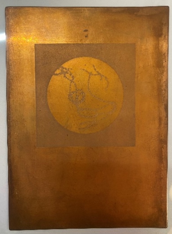
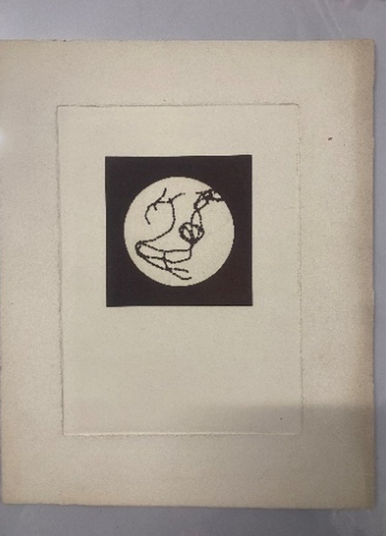
On the left, the copper plate; on the right, the corresponding photogravure print. The image is reversed on printing.
 If you are interested in the museum project or have technical or scientific objects that can provide clues to the past activities of the Institut Pasteur's laboratories, feel free to contact the museum team: musee@pasteur.fr